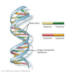
If you remember your high school biology, you know that DNA has a "double helix" structure. It made up of pairs of proteins (Adenine/Thymine and Guanine/Cytosine) with a sugar molecule on one side and a phosphate molecule on the other. When you stack them on top of each other, it forms the spiraling "helix" structure we think of for DNA. All of this is old news. It was discovered by Watson and Crick ( well, really Rosalind Franklin) back in the 50's.
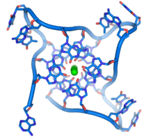
What is new to me, is that DNA can also form into a quadruple helix. In this case, four molecules of a single nucleic acid (ie Guanine) bond together to make a flat plate structure. When these plates stack on top of each other you get a "G-quadraplex" aka quadruple helix DNA. However, historically, this was just a fun lab trick. There was no evidence that such a structure actually formed in nature in any stable way.
But a recent report in Nature Chemistry, shows evidence that the G-quadraplexes are present in cancer cells. New Scientist interviewed one of the researchers who explained that the G-quadraplexes are typically temporary structures present during the division of cells and are present in a structure called the telemere, the cap of a chromosome. Since cancer cells typically replicate very quickly, telemeres are frequently not replicated accurately.
What is really exciting is that the compound the team used to stabalize the G-quadraplexes in cancer cells and prevent them from naturally reverting to a double helix (pyridostatin) has been previously shown to prevent replication of breast cancer cells. This result suggests that pyridostatin could potentially be used to treat cancer and that further understanding of the transitory nature of DNA structures is an important thing to understand.
Score one for playing around in the lab. Off to light things on fire and see what color they burn.
Image Credits: NIH, Wikipedia
 RSS Feed